
Fig. 5
- ID
- ZDB-FIG-170620-7
- Publication
- Miller et al., 2017 - A genetic basis for molecular asymmetry at vertebrate electrical synapses
- Other Figures
- All Figure Page
- Back to All Figure Page
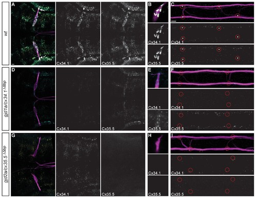
Recruitment of Gjd1a/Cx34.1 and Gjd2a/Cx35.5 to electrical synapses is co-dependent. Larvae are stained for anti-GFP (magenta), anti-Connexin34.1 (Cx34.1, yellow), and for anti-Connexin35.5 (Cx35.5, cyan). Individual Cx34.1 and Cx35.5 channels are shown in neighboring panels. Hindbrain, lateral dendrite, and spinal cord images are maximum intensity projections of ~30,~5, and ~10 uM, respectively. Anterior is to the left. Scale bar = 10 uM. (A–C) Cx34.1 and Cx35.5 are found colocalized at electrical synapses throughout the hindbrain (A) including at Aud/M (arrows in A, (B), and M/CoLo synapses (C). In gjd1a/cx34.1 mutants the localization of Cx35.5 to electrical synapses is lost (D–F) or diminished at Aud/M synapses (E); in gjd2a/cx35.5 mutants the localization of Cx34.1 to electrical synapses is lost (G–I). Associated antibody information can be found in source data for Figure 5. |
| Gene: | |
|---|---|
| Antibodies: | |
| Fish: | |
| Anatomical Terms: | |
| Stage: | Day 5 |
| Fish: | |
|---|---|
| Observed In: | |
| Stage: | Day 5 |