
Fig. 4 S1
- ID
- ZDB-FIG-170609-22
- Publication
- Venero Galanternik et al., 2017 - A novel perivascular cell population in the zebrafish brain
- Other Figures
- All Figure Page
- Back to All Figure Page
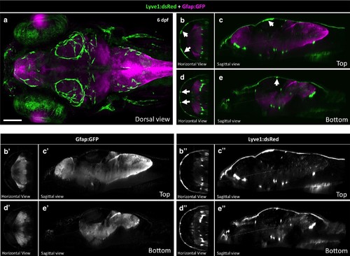
Radial Glia and FGPs localize to different parts of the brain. (a) Dorsal view of a Z-projected confocal image generated by the Zebrafish Brain Browser (Marquart et al., 2015) superimposing four 6 dpf Tg(lyve1:dsRed) (green) embryos to Tg(gfap:GFP) (magenta). Rostral is to the left. (b,d) Horizontal views of panel A at the top (b) and bottom (d) of the Z-stack across the entire brain. (c,e) Sagittal views of panel A at the top (c) and bottom (e) Z-sections. White arrows in panels b-e show FGPs. Panels b'-e' show the Gfap:GFP channel only. Panels b'-e' show the Lyve1:dsRed channel only. Scale bar for all panels: 100 μm. |