
FIGURE
Fig. 2 S1
- ID
- ZDB-FIG-170609-19
- Publication
- Venero Galanternik et al., 2017 - A novel perivascular cell population in the zebrafish brain
- Other Figures
- All Figure Page
- Back to All Figure Page
Fig. 2 S1
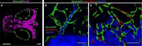
Mrc1a-positive perivascular cells are EdU negative and fail to collect and drain dye like Mrc1a-positive lymphatics. (a) EdU staining (magenta) of a 5 dpf Tg(mrc1a:eGFP) transgenic embryo where Mrc1a:eGFP-positive cells (green) are not EdU positive (n = 5 animals). Rostral is to the left. (b) Lymphatic vessel (green) on the head of a juvenile Tg(mrc1a:eGFP) transgenic animal showing lymphatic drainage of injected Qdots-705 (blue). (c) FGPs (green) on the brain surface of a Tg(mrc1a:eGFP) transgenic juvenile do not drain injected Qdots-705 (blue). n = 2 injected fish. Scale bars: 100 µm. |
Expression Data
Expression Detail
Antibody Labeling
Phenotype Data
Phenotype Detail
Acknowledgments
This image is the copyrighted work of the attributed author or publisher, and
ZFIN has permission only to display this image to its users.
Additional permissions should be obtained from the applicable author or publisher of the image.
Full text @ Elife