
Fig. 1
- ID
- ZDB-FIG-170530-4
- Publication
- Chow et al., 2017 - In Vivo Measurement of Glycine Receptor Turnover and Synaptic Size Reveals Differences between Functional Classes of Motoneurons in Zebrafish
- Other Figures
- All Figure Page
- Back to All Figure Page
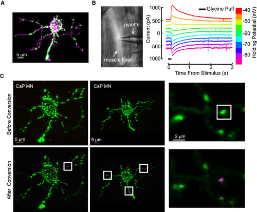
The Expression of dendra2-GlyRα1 in Motoneurons, Physiology of dendra2-GlyRα1 Channels in Muscle Fibers, and Synapse Level Targeting for dendra2 Photoconversion (A) A single motoneuron expressing dendra2-GlyRα1 (green) along with membrane targeted tdTomato (magenta), exhibiting distributed glycinergic puncta along the labeled soma and dendrites. See also Figure S1 for costaining with gephyrin. (B) Expression of dendra2-GlyRα1 in muscle to test channel formation. Left: transgenic muscle fiber targeted for patch recording. Right: voltage-clamp recordings at the indicated holding potentials in a muscle fiber. (C) Precise targeting with a 405-nm laser allowed us to convert synaptically localized dendra2-GlyRα1 from green (top) to red fluorescence (bottom, represented as magenta) with near single-synapse accuracy. |