
Fig. 2
- ID
- ZDB-FIG-170523-33
- Publication
- Thomas-Jinu et al., 2017 - Non-nuclear Pool of Splicing Factor SFPQ Regulates Axonal Transcripts Required for Normal Motor Development
- Other Figures
- All Figure Page
- Back to All Figure Page
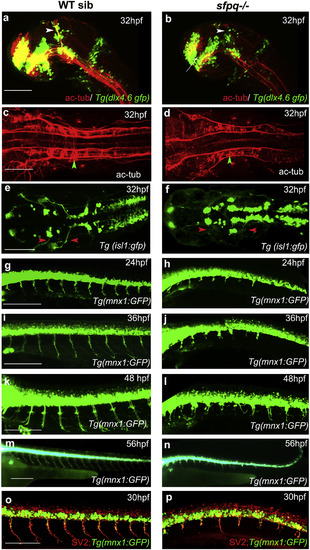
Axonogenesis Is Affected in sfpq−/− Embryos Lateral (A, B, and G–P) and dorsal (C–F) views, anterior to the left, of zebrafish brain at 32 hpf (A and B) and spinal cord at 24–56 hpf. (A and B) sfpq;Tg(dlx4.6:gfp) embryos. Lack of supra-optic commissure (white arrow) and posterior commissure (white arrowhead) in coma (n = 8/32) is revealed by acetylated tubulin staining. (C and D) Dorsal view, anterior to the left of acetylated tubulin staining showing hindbrain disorganized axonal tracks in sfpq mutant (D; green arrowhead, n = 8) compared to siblings (C; n = 26). (E and F) Dorsal view, anterior to the left of GFP+ motor neurons in sfpq; Tg(isl1:gfp) siblings (E) and mutant (F), showing cranial motor neuronal clusters lacking axonal projections in the mutant (red arrowhead, n = 7). (G–N) Lateral view, anterior to the left, of confocal live imaging, showing temporal defect in axonogenesis in the majority of spinal motor neurons in a mutant (H, J, L, and N; n = 14) compared to a sibling (G, I, K, and M; n = 42) in the Tg(mnx1: gfp) background. (O and P) Lateral view, anterior to the left, of SV2 antibody staining of siblings (O) and sfpq null mutant (P), showing pre-synaptic protein in the few axons formed in sfpq mutants (n = 9). Scale bar, 100 μm. |
| Fish: | |
|---|---|
| Observed In: | |
| Stage Range: | Prim-5 to Long-pec |