
Fig. S7
- ID
- ZDB-FIG-170523-29
- Publication
- Lopez et al., 2017 - A152T tau allele causes neurodegeneration that can be ameliorated in a zebrafish model by autophagy induction
- Other Figures
- All Figure Page
- Back to All Figure Page
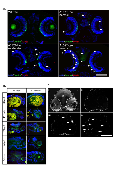
Cell death in Dendra-tau zebrafish Further data supporting main Fig.4. (A) Representative images of TUNEL labelling in brain sections showing consistent and reproducible differences in cell death between WT- and A152T-tau fish according to morphological phenotype (normal, moderate and severe) at 6 d.p.f. Apoptotic nuclei are highlighted by white arrowheads and quantification is shown in main Fig. 4E. B) TUNEL labelling on longitudinal sections of WT- and A152T-tau fish from 24 h.p.f. to 5 d.p.f. used to identify the time points at which cell death is occurring. The timecourse showed a larger number of apoptotic cells in mutant A152T-tau compared to WTtau fish in all ages, most evident at 2 d.p.f. (C) TUNEL labelling in brain sections of WT- and A152T-tau fish at 2 d.p.f. representative of those used for quantification of the number of apoptotic cells presented in main Fig.4D (l=lens; r=retina and b=brain) . A-C scale bar represents 100 μm. |