
Fig. 5
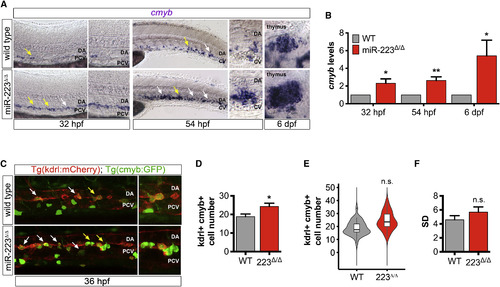
miR-223 Does Not Regulate Phenotypic Variability of HSPC Number (A) WISH of HSPC marker cmyb at transient sites of hematopoiesis, namely the DA ventral wall at 32 hpf, the caudal hematopoietic tissue between the DA and CV at 54 hpf, and the thymus, a definitive site of hematopoiesis, at 6 dpf (n = 10–20 embryos). Arrows show examples of cmyb+ cells. Yellow arrows indicate the region of the lateral trunk that is depicted in magnified images. Images captured with a 20× objective. (B) Mean cmyb expression in miR-223Δ/Δ embryos normalized to actb1 levels and compared with wild-type as determined by qRT-PCR for three biological replicates. Bar plots show mean ± SEM, two-tailed Student's t test. (C) Representative micrographs (25×) of the lateral trunk in embryos expressing Tg(kdrl:ras-mCherry)s896 and Tg(cmyb:GFP)zf169. Arrows point to examples of kdrl+cmyb+ cells budding from the DA ventral wall at the peak of hematopoiesis. Yellow arrows show region of the lateral trunk that is captured in zoomed-in images. (D) Bar plots show the average of replicate means ± SEM of kdrl+cmyb+ cell number at 36 hpf (n = 4 replicates). (E) Violin plots show kdrl+cmyb+ cell number probability density distributions. Solid lines in box plots depict median values. Phenotypic variability was not significantly different from wild-type, p = 0.24 by Levene's test (n = 36 embryos). (F) Bar plots show average replicate SD ± SEM of kdrl+cmyb+ cell number (n = 4 replicates). Significance calculations were relative to wild-type embryos. n.s., not significant (p > 0.05); ∗p ≤ 0.05, ∗∗p ≤ 0.01, two-tailed Mann-Whitney U test unless otherwise indicated. Abbreviations as defined in Figure 1. |
| Genes: | |
|---|---|
| Fish: | |
| Anatomical Terms: | |
| Stage Range: | Prim-15 to Day 6 |
| Fish: | |
|---|---|
| Observed In: | |
| Stage Range: | Prim-15 to Day 6 |
Reprinted from Developmental Cell, 40, Kasper, D.M., Moro, A., Ristori, E., Narayanan, A., Hill-Teran, G., Fleming, E., Moreno-Mateos, M., Vejnar, C.E., Zhang, J., Lee, D., Gu, M., Gerstein, M., Giraldez, A., Nicoli, S., MicroRNAs Establish Uniform Traits during the Architecture of Vertebrate Embryos, 552-565.e5, Copyright (2017) with permission from Elsevier. Full text @ Dev. Cell