|
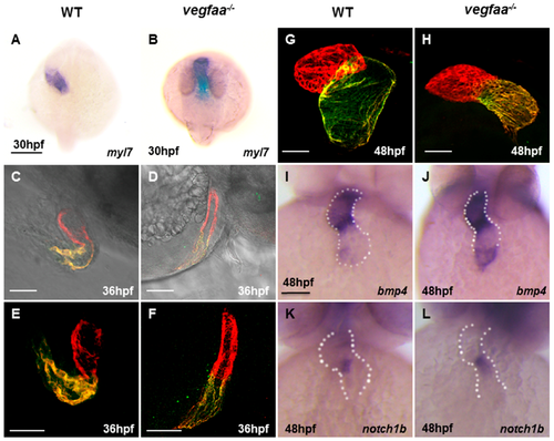
vegfaa deficiency results in defects in cardiac looping and chamber formation. (A,B) Dorsal views of in situ hybridization depicts myl7 expression at 30 hpf in wild-type embryos (A) and vegfaa mutant embryos (B). Scale bar: 200 μm (A,B); (C–F) Lateral views of zebrafish hearts stained with MF20 (red) and S46 (green) antibodies to visualize the ventricle and atrium at 36 hpf. MF20 marks the whole heart, whereas S46 is atrium-specific. Scale bar: 50 μm. (C,D) are added with light microscopy; (G–H) At 48 hpf, the wild-type heart is looped, with morphologically distinct chambers, whereas the vegfaa mutant heart appears unlooped, with a small heart, especially ventricle. Scale bar: 50 μm (C– H); (I–L) Frontal views depict expression of the atrio-ventricular canal (AVC) markers in wild-type (I,K) and in vegfaa mutants (J,L) at 48 hpf. Dotted lines outline the chambers flanking the AVC. Scale bar: 100 μm (I–L).
|