
Fig. S7
- ID
- ZDB-FIG-170315-34
- Publication
- Ye et al., 2015 - Endoderm convergence controls subduction of the myocardial precursors during heart-tube formation
- Other Figures
- All Figure Page
- Back to All Figure Page
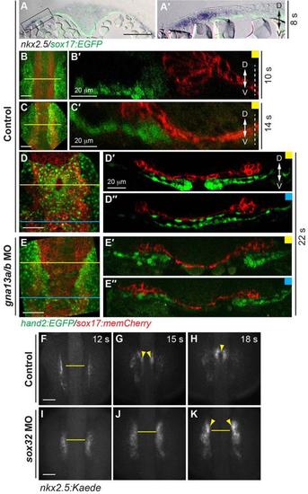
The ALPM engages in subduction, with some cells migrating from the dorsal to the ventral side of the endoderm. (A) The relative positions of the ALPM and endoderm. (A-A') Overlays of GFP (endoderm) and hand 2 expression as detected by ISH in Tg(sox17:EGFP) embryos at 8s. (A'): high-magnification image of area indicated in A. (B-E'') Confocal images of control and gna13a/b MO-injected Tg(hand2:EGFP)/Tg(sox17:memCherry) embryos at the indicated stages, showing the EGFP-expressing ALPM and memCherry-expressing endoderm. (B-E) Z-projections. Horizontal lines: positions of the XZ sections. (B'-E'') XZ views of z-stacks of embryos at positions indicated in B-E. White dashed lines: midline. D: dorsal; V: ventral. (F-K) Snapshots of myocardial precursors from epifluorescence time-lapse movies taken of control (F-H) or sox32 MO-injected (I-K) Tg(nkx2.5:Kaede) embryos at the indicated stages, using a 5x/NA 0.15 objective. Yellow arrowheads: myocardial cells; yellow lines: the distance between the two populations of myocardial cells (equivalent length). Scale bars: 100 μm, unless stated otherwise. |