
Fig. 1
- ID
- ZDB-FIG-170125-27
- Publication
- Willis et al., 2016 - Injections of Predatory Bacteria Work Alongside Host Immune Cells to Treat Shigella Infection in Zebrafish Larvae
- Other Figures
- All Figure Page
- Back to All Figure Page
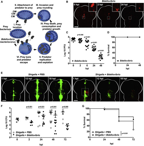
Injected Predatory Bdellovibrio Persist in Zebrafish Larvae without Ill Effects and Protect against Shigella Infection In Vivo (A) Cartoon of Bdellovibrio life cycle. (I–III) Motile predatory Bdellovibrio attach to and invade the periplasm of Gram-negative bacteria such as Shigella. (III) Prey bacteria are rounded by DD-endopeptidase action on the cell wall. (IV) Prey bacteria are killed in ∼30 min and kept intact as Bdellovibrio consume their contents and grow. (V and VI) Following replication, Bdellovibrio lyse prey 180–240 min after invasion, releasing further predators. These Bdellovibrio progeny can repeat the predatory cycle. OM, outer membrane; CW, cell wall peptidoglycan; CM, cytoplasmic membrane. (B) Wild-type (WT) AB larvae were injected at 3 dpf in the hindbrain ventricle with 1–10 × 104 PFUs of mCherry-Bdellovibrio (red). The same larvae were imaged over time to observe distribution. Representative images from a single larva are shown here. Scale bar, 100 μm. (C) Enumeration of live Bdellovibrio in PBS-homogenates from larvae injected with mCherry-Bdellovibrio as in (B) over time. Each circle represents a count from an individual larva. Data are pooled from two independent experiments (n = 8 larvae per experiment). Mean ± SEM (horizontal bars) is shown. The p values (versus the 0 hpi time point) were determined by multiple t test. Significance with Bonferroni correction was defined as p < 0.0125. See also Figures S1B–S1D for comparative evaluations of Bdellovibrio persistence from different doses in larvae at different developmental stages. (D) Survival curve of WT AB larvae injected with mCherry-Bdellovibrio as in (B) and incubated at 28°C for 48 hpi. Data are pooled from three independent experiments (n = 22–37 larvae per experiment). (E) WT AB zebrafish larvae were injected in the hindbrain ventricle at 3 dpf with >5 × 10³ CFUs of GFP-S. flexneri (green), followed by a hindbrain injection of either PBS or 1–2 × 10⁵ PFUs of mCherry-Bdellovibrio (red), 30–90 min after the initial Shigella infection. Representative images of the hindbrain ventricle in PBS- or Bdellovibrio-treated zebrafish larvae infected with Shigella are shown. Dotted square shows region of interaction between fluorescent Shigella and Bdellovibrio. For each treatment, the same larva was imaged over time. Scale bar, 100 μm. See also Movie S1. (F) Enumeration of live Shigella in homogenates of larvae injected with S. flexneri and treated with injections of either PBS or Bdellovibrio as in (E) over time. Each circle represents a count from an individual larva. Half-filled circles represent enumerations from larvae at time 0 and are representative of inocula for both conditions. Only viable larvae were included in the analysis. Data are pooled from four independent experiments (up to n = 3 larvae per time point per experiment). Mean ± SEM (horizontal bars) is shown. The p values (between conditions at cognate time points) were determined by unpaired one-tailed Student’s t test. Significance was defined as p < 0.05. (G) Survival curve of larvae injected with S. flexneri and treated with either PBS or Bdellovibrio as in (E). Larvae were incubated at 28°C for 72 hpi. Data are pooled from three independent experiments (n = 22–48 larvae per condition per experiment). Up to three larvae per condition were taken for CFUs at 2, 24, 48, and 72 hr time points. The p value between conditions was determined by log-rank Mantel-Cox test. Significance was defined as p < 0.05. See also Figure S1. |