
Fig. 3
- ID
- ZDB-FIG-170110-26
- Publication
- Xiao et al., 2016 - Chromatin-remodelling factor Brg1 regulates myocardial proliferation and regeneration in zebrafish
- Other Figures
- All Figure Page
- Back to All Figure Page
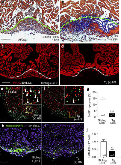
Inhibition of brg1 impairs cardiac regeneration. (a–d) Representative sections from wild-type siblings (a,c) and Tg(hsp70:dn-xbrg1) (b,d) hearts at 30 d.p.a., evaluated by AFOG staining (a,b), and immunofluorescence staining with anti-myosin heavy chain (MF20) (c,d). Note massive fibrosis (b) and compromised myocardial regeneration (d) in Tg(hsp70:dn-xBrg1) hearts (tg). Dashed lines mark the resection site. (e–g) Paraffin sections of 14 d.p.a. regenerating heart of wild-type sibling (e) and Tg(hsp70:dn-xBrg1) (f) hearts co-stained for BrdU (green), Mef2C (red) and 4,6-diamidino-2-phenylindole (DAPI; blue). Higher-magnification images of areas in squares are shown in the upper-right corners, and Mef2C+/BrdU+ double-positive cardiomyocytes are indicated by arrowheads. (g) Percentages of Mef2C+/BrdU+ cardiomyocytes in the injured area (***P<0.001; n=6 for siblings and 7 for transgenic hearts; data are mean percentages±s.e.m., paired Student’s t-test). (h–j) Paraffin sections of 14 d.p.a. wild-type Tg(gata4:EGFP) sibling (h) and Tg(hsp70:dn-xbrg1; gata4:EGFP) (i) hearts stained with anti-EGFP and DAPI. The average of fluorescence intensity was calculated using Imaris software (j) (**P<0.01; n=6; data are mean percentages±s.e.m.; paired Student’s t-test). Scale bars, 100 μm. |