
Fig. S6
- ID
- ZDB-FIG-170104-20
- Publication
- Jiménez-Amilburu et al., 2016 - In Vivo Visualization of Cardiomyocyte Apicobasal Polarity Reveals Epithelial to Mesenchymal-like Transition during Cardiac Trabeculation
- Other Figures
- All Figure Page
- Back to All Figure Page
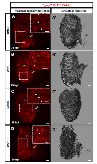
DAPT-treated hearts form disorganized trabecular ridges (A-D') 3D spinning disk images and surface renderings at mid-sagittal plane of 79 (A-B) and 98 (C-D) hpf Tg(myl7:MKATE-CAAX) zebrafish hearts. Dashed boxes show higher magnification images. DMSO-treated larvae show a smooth cardiac wall and clear trabecular ridges in the cardiac lumen at 79 (A and A', arrowheads) and 98 (C and C', arrowheads) hpf, while larvae treated with 100 μM DAPT starting at 48 hpf show a collapsed cardiac wall and disorganized trabecular structures at 79 (B and B', asterisks) and 98 (D and D', asterisks) hpf. V, ventricle; AVC, atrioventricular canal. Scale bars, 10 μm. |