
Fig. 7
- ID
- ZDB-FIG-170104-14
- Publication
- Jiménez-Amilburu et al., 2016 - In Vivo Visualization of Cardiomyocyte Apicobasal Polarity Reveals Epithelial to Mesenchymal-like Transition during Cardiac Trabeculation
- Other Figures
- All Figure Page
- Back to All Figure Page
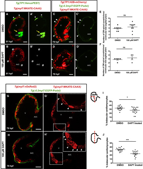
Blocking Notch Activation Did Not Increase the Number of CMs Exhibiting Apical Constriction or of CMs in the Trabecular Layer (A–B') Confocal images (mid-sagittal sections) of 81-hpf Tg(myl7:MKATE-CAAX);Tg(TP1:VenusPEST) zebrafish hearts. DMSO-treated larvae show Notch activity in CMs (A and A', asterisks) and atrioventricular canal (AVC) cells (A and A', yellow arrows). Larvae treated with 100 μM DAPT lack Notch activity in both CMs and AVC cells (B and B', Ø). (C–D') Confocal images (mid-sagittal sections) of 79-hpf Tg(−0.2myl7:EGFP-podxl);Tg(myl7:MKATE-CAAX);Tg(TP1:H2B-mCherry) zebrafish hearts. DMSO-treated larvae show CM apical constrictions (C and C', arrowheads). 79-hpf larvae treated at 48 hpf with 100 μM DAPT show similar numbers of CM apical constrictions than controls (D and D', arrowheads). (E and F) Number of CM apical constrictions per ventricle at the mid-sagittal plane after 100 μM DAPT treatment. Animals were treated at 48 hpf and imaged at 79 hpf (E) or treated at 60 hpf and imaged at 72 hpf (F). Each dot represents one heart. Data are shown as mean ± SEM. ns, no significant differences by Student’s t test. (G–H') Confocal images (mid-sagittal sections) of 79-hpf Tg(−0.2myl7:EGFP-podxl);Tg(myl7:MKATE-CAAX);Tg(myl7:nDsRed2) zebrafish hearts. DMSO-treated larvae show normal CM shape in the compact layer (G', arrowheads), while DAPT-treated larvae show abnormal CM shape (H', arrowheads). (I and J) Graphs showing the percentage of trabecular CMs versus the total number of CMs counted in two regions of the outer curvature. Each dot represents one heart. Data are shown as mean ± SEM. ∗p < 0.05 and ∗∗∗p < 0.001 by Student’s t test. V, ventricle; AVC, atrioventricular canal. Scale bars, 20 μm. |
| Genes: | |
|---|---|
| Fish: | |
| Condition: | |
| Anatomical Terms: | |
| Stage: | Protruding-mouth |
| Fish: | |
|---|---|
| Condition: | |
| Observed In: | |
| Stage: | Protruding-mouth |