
Fig. 5
- ID
- ZDB-FIG-170104-12
- Publication
- Jiménez-Amilburu et al., 2016 - In Vivo Visualization of Cardiomyocyte Apicobasal Polarity Reveals Epithelial to Mesenchymal-like Transition during Cardiac Trabeculation
- Other Figures
- All Figure Page
- Back to All Figure Page
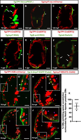
Notch-Signaling-Reporter-Positive CMs Appear in the Compact Layer Adjacent to CMs Undergoing Apical Constriction (A–C) Confocal images (mid-sagittal sections) of Tg(myl7:ras-mCherry);Tg(TP1:VenusPEST) zebrafish larvae at 76 (A), 100 (B), and 125 (C) hpf. Asterisks indicate CMs expressing the Notch signaling reporter. White arrows in (A) point to fading TP1:VenusPEST expression in the endocardium. Yellow arrows point to the developing AV valve in (A) and (B). (D–F) Confocal sections of 7-dpf larval hearts obtained from crossing the Tg(TP1:CreERT2) line to three different indicator lines. In each case, embryos were treated with 4-OHT from 2 to 5 dpf. Asterisks indicate labeled CMs in the compact layer. (D) Lineage-traced 7-dpf Tg(TP1:CreERT2);Tg(myl7:RSG) heart. Arrowhead points to a CM neighboring the AV canal. (E) Lineage-traced 7 dpf Tg(TP1:CreERT2);Tg(β-act2:RSG) heart. (F) Lineage-traced 7 dpf Tg(TP1:CreERT2);Tg(ubi:Switch) heart. Note that in this case, lineage-traced cells are red. White arrows point to labeled endocardial cells. (G–J') Confocal images (mid-sagittal sections) of 79-hpf Tg(−0.2myl7:EGFP-podxl);Tg(myl7:MKATE-CAAX);Tg(TP1:H2B-mCherry) zebrafish hearts. Boxed areas in (G) and (H) are shown in G' and H', respectively. Notch-reporter-positive CMs (G' and H', asterisks) localize in the compact layer adjacent to CMs undergoing apical constriction (G' and H', white arrowheads). Boxed areas in (I) and (J) are shown in (I') and (J'), respectively. Notch-reporter-positive CMs (I' and J', asterisks) appear directly adjacent to a delaminated CM (I' and J', //). Purple arrowheads point to Notch-reporter-positive endocardial cells (G', H', I', and J'). (K) Graph shows percentage of CMs exhibiting apical constriction that are surrounded by Notch-reporter-positive CMs per heart at 79 hpf. Each dot represents one heart. At, atrium; V, ventricle; AVC, atrioventricular canal. Scale bars, 20 μm. |
| Genes: | |
|---|---|
| Fish: | |
| Condition: | |
| Anatomical Terms: | |
| Stage Range: | Protruding-mouth to Days 7-13 |