
Fig. 3
- ID
- ZDB-FIG-161220-9
- Publication
- Greaney et al., 2017 - Extraocular motoneuron pools develop along a dorsoventral axis in zebrafish, Danio rerio
- Other Figures
- All Figure Page
- Back to All Figure Page
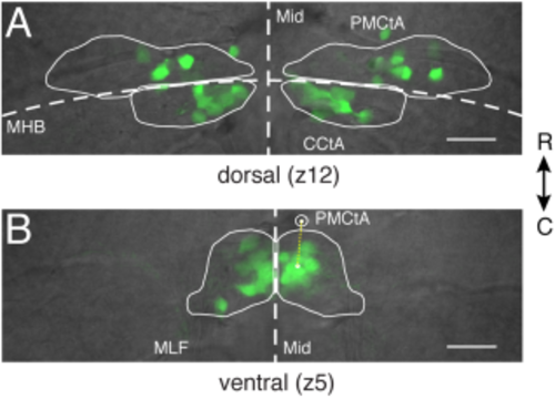
Fluorescent somata in Tg(isl1:GFP) zebrafish define the spatial extent of extraocular motoneurons in nIII and nIV. (A) Positions of fluorescent motoneurons in dorsal nIII/nIV (z12) relative to anatomical landmarks. nIII: rostral to MHB; nIV: caudal to MHB. (B) Positions of fluorescent motoneurons in ventral nIII (z5) relative to anatomical landmarks. Yellow dotted line represents the distance between the center of intensity of green fluorescence right of the midline and the centroid of the right PMCtA. Mid: midline. MHB: midbrain–hindbrain boundary. MLF: medial longitudinal fasciculus. PMCtA: posterior mesencephalic central artery. CCtA: cerebellar central artery. Scale bars = 20 μm. |