
Fig. 2
- ID
- ZDB-FIG-161220-8
- Publication
- Greaney et al., 2017 - Extraocular motoneuron pools develop along a dorsoventral axis in zebrafish, Danio rerio
- Other Figures
- All Figure Page
- Back to All Figure Page
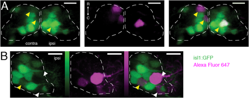
Identification of dye-filled motoneuron subtypes by projection pattern and Tg(isl1:GFP) expression. (A) Filled motoneurons in ventral nIII (plane z4, or 5th of 15 6-μm-thick planes counting dorsally from z0, dashed line). Yellow arrowheads indicate dye-filled motoneurons that were also GFP+ (contralateral: SR motoneurons; ipsilateral: IR/MR motoneurons). (B) Filled motoneurons in ventral nIII (plane z5, dashed line). White arrowheads indicate dye-filled motoneurons without GFP (IO motoneurons); yellow arrowhead indicates a dye-filled motoneuron that was also GFP+ (IR/MR motoneuron). Color bars represent photons detected in photon-counting mode (IO data only); green range: 0–198; magenta range: 0–231. Green = GFP; magenta = Alexa Fluor 647 dye. Scale bars = 10 μm. |