
Fig. 4
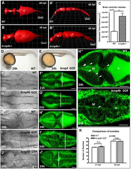
Kcng4b regulates the brain ventricular system. (A,B) 3D reconstruction of confocal Z-scans of the brain ventricular system in vivo after injection with 70 kDa Texas Red-Dextran in dorsal view (A,A′) and tilted lateral view (B,B′). (C) Volume measurement of the brain ventricular system by 3D-reconstuction of Z-scans sections using surface function of Imaris 7.0. Data represent mean±s.e.m. of n=7; **P<0.01, unpaired t-test. (D-J) kcng4b GOF 21-24 hpf embryos. (D,E) Morphology of embryos (lateral view). (F-J) Dorsal view of embryos labelled in vivo by FITC-BODIPY-ceramide at 21-24 hpf, confocal DIC and maximal projection of Z-scans. The perpendicular line marks the widest opening of the hindbrain ventricle. (H″,I″) Confocal plane showing the ventricle with BODIPY-labelled cell membranes. Note that the rounded cells facing the lumen (arrowhead) are less abundant in kcng4b GOF (I″) compared with control (H″). (K) Number of somites at 21 and 24 hpf. Data represent mean±s.e.m.; n=25; n.s., not significant; unpaired t-test. Scale bars: 100 µm (A-B′), 50 µm (D-J). |
| Fish: | |
|---|---|
| Observed In: | |
| Stage: | Long-pec |