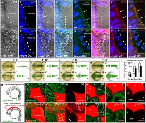
Analyses of tissue integrity. (A-H′) Confocal sections (dorsal view), in the top left corner of the anterior portion of the third ventricle. Controls (A-H) and kcng4b morphants (A′-H′). In the WT control, the apical membrane (arrow) is continuous (A); in the morphants, it is discontinuous with cells in the ventricular cavity (A′). (B′,C′) DAPI stains cell nuclei (arrowhead) in the ventricular lumen (dashed line) of kcng4b morphants. (D-H′) Apical markers ZO-1 (green, D-E′) and aPKC (red, F-G′) clearly define plasma membrane in WT, but not in kcng4b morphants. (E,E′,G,G′) Zoomed imaged of boxed areas in D,D′ and F,F′, respectively. (H,H′) Merged image from E,G and E′,G′, respectively. Images are whole mount 24 hpf embryos (dorsal view) taken with a 63× water dipping objective. (I-P′) Dye retention assay in WT and kcng4b morphants (24 hpf). Blue brackets in K,L indicate dye front. (Q) Dye leakage was quantified by measuring the extent of migration of the dye front along the blue line in K and L. Each time point represents an average of data from six independent experiments. Error bars are s.e.m. *P<0.05, **P<0.01 compared with control (unpaired Student's t-test). (R) Confocal analysis of 70 kDa Dextran leakage was performed after injection into fourth ventricle. (S-Z) Confocal section (dorsal view) of neuroepithelium 30 min after dye injection into wild-type and kcng4b MO embryo in Tg(h2b:EGFP) background at 24 hpf. Dye is seen in forebrain neuroepithelium of kcng4b MO embryos (arrows in V,V′; arrowheads show delaminated cells in V′) but not in midbrain and hindbrain neuroepithelium (X,Z). U-V′ and Y-Z′ are zoomed images of boxed areas in S,T and W,X, respectively. H, hindbrain; M, midbrain; MHB, midbrain-hindbrain boundary; ventricles are labelled with roman numerals. Scale bars: 10 µm (A-P); 25 µm (S-Z).
|
