
Fig. S2
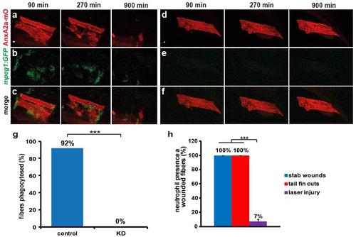
Macrophages remove severely damaged myofibers. a-f, Dead myofibers show precipitation of AnxA2a-mO (red) to internal membranes (a and d). In controls dead myofibers were removed by attracted macrophages (b, green) within 900 min after wounding (c). In morphants dead myofibers persisted beyond imaging time (d and f), due to the absence of macrophages (e). Panels a-b and d-e were merged in c and f, respectively. g, Macrophages remove dead myofibers. In controls (left, n=12), 92% of dead myofibers were removed by macrophages within 20 h of observation. In contrast, removal of dead cells (right, n=20) was totally abolished (0%) in animals lacking macrophages. h, Neutrophils were barely recruited to wounds inflicted with a laser (purple, n=126) while stab wounds caused by insertion of a glass needle into the somitic musculature (blue, n=23) and cutting of the tail fin (red, n=12) attracted neutrophils in all cases. Thus infection and concomitant differences in inflammatory status may have a bearing on which immune cells become engaged. Data was analyzed as maximum projections. p-values were calculated according to Fisher’s exact test, p<0.001. Scale bars: 4 µm. |