
Fig. S1
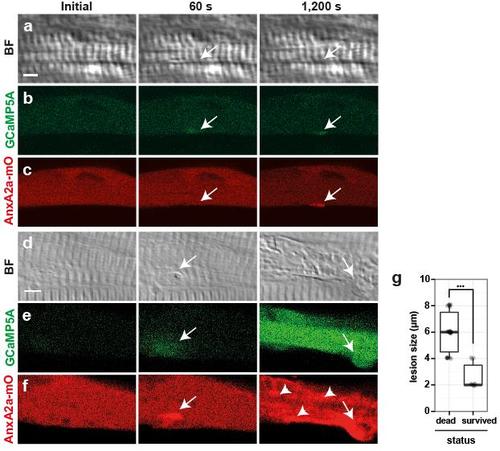
Fate of myofibers depends on size of membrane wound. a-f, Myofibers with a lesion (arrows) of either ≤4 µm (a-c) or ≥4 µm (d-f) were analyzed for repair patch formation (AnxA2a-mO, c, f), local Ca2+ changes (GCaMP5A, b, e) and corresponding bright field views (BF, a, d), prior to (initial), 60 s and 1,200 s after wounding. Immediately after wounding, GCaMP5A was locally activated regardless of the lesion size (b, e). Ca2+ levels at lesions of ≤ 4 µm size resolved back to baseline after 1,200 s with the exception of Ca2+ in the repair patch (b), however, lesions ≥4 µm led to propagation of a Ca2+ wave across the entire cell (e), frequently followed by sliding of the repair patch along the myofiber (arrow, d-f), and precipitation of AnxA2a-mO to internal membranes of myofibers (f, arrowheads), a hallmark of cell death. g, Survival rate of myofibers was analyzed as a function of initial lesion size (Y-axis in µm). Myofibers with lesions ≥4 µm underwent cell death (left column; n=30), while cells with lesions ≤4 µm survived (right column: n=10; Student’s t-test, p<0.001). Scale bar: 4 µm. |