
Fig. 3
- ID
- ZDB-FIG-161019-6
- Publication
- Ellis et al., 2016 - Experimental Dissection of Metalloproteinase Inhibition-Mediated and Toxic Effects of Phenanthroline on Zebrafish Development
- Other Figures
- All Figure Page
- Back to All Figure Page
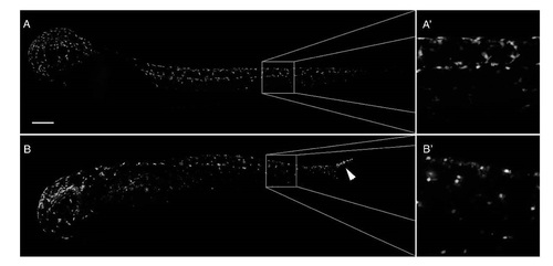
PhN perturbs development of neural crest-derived pigment cells. (A) A 48 hpf Tg(mitfa:eGFP) embryo exposed from 24 hpf to vehicle control, exhibiting normal distribution of GFP-expressing presumptive pigment cells. At high magnification (A′), the stellate morphology of these cells is apparent as they invade the overlying epidermis; (B) A 48 hpf Tg(mitfa:eGFP) embryo exposed from 24 hpf to 10 µM PhN, in which presumptive pigment cells in the head and anterior trunk have successfully emigrated from the neural tube and become distributed relatively normally, but in which presumptive pigment cells in the posterior tail have failed to emigrate from the neural tube (arrowhead). At high magnification (B′), these cells have a rounded amoeboid appearance. Boxes indicate regions magnified. Scale bar = 200 µm. |