
FIGURE
Fig. 7
- ID
- ZDB-FIG-161019-10
- Publication
- Ellis et al., 2016 - Experimental Dissection of Metalloproteinase Inhibition-Mediated and Toxic Effects of Phenanthroline on Zebrafish Development
- Other Figures
- All Figure Page
- Back to All Figure Page
Fig. 7
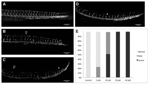
PhN disrupts angiogenesis. Forty-eight hpf Tg(fli1:eGFP) embryos treated from 24 to 48 hpf with either: (A) vehicle alone; or various concentrations of PhN (B-D), illustrating: (B) "Normal"; (C) "Mild"; and (D) "Severe" disruption of angiogenesis. The DLAV is indicated with an arrowhead in (B,C); and examples of disrupted angiogenesis are highlighted with asterisks; (E) Summary of blind scoring of 40 embryos exposed to vehicle alone, 96 embryos exposed to 5 µM PhN, 102 embryos exposed to 10 µM PhN, 91 embryos exposed to 25 µM PhN, and 77 embryos exposed to 40 µM PhN. |
Expression Data
Expression Detail
Antibody Labeling
Phenotype Data
Phenotype Detail
Acknowledgments
This image is the copyrighted work of the attributed author or publisher, and
ZFIN has permission only to display this image to its users.
Additional permissions should be obtained from the applicable author or publisher of the image.
Full text @ Int. J. Mol. Sci.