
Fig. S2
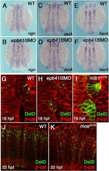
Epb41l5 does not modify Notch signaling or subcellular distribution of DeltaD in the neuroepithelium. (A-F) Reduced expression of epb41l5 does not change fate specification of neurons mediated by Notch-mediated lateral inhibition. ngn: bHLH proneural transcription factor, delta: Notch ligand, her4: direct outcome of Notch signaling. 3-somite stage embryos. (G-I) Reduced expression of Epb41l5 does not change subcellular distribution of DeltaD in the hindbrain neuroepithelium. Dorsal views of the 4th rhombomere and the otic placode in 10-somite stage embryos. DeltaD is mainly localized in cytoplasmic puncta in wild-type embryos. DeltaD is mainly localized on the plasma membrane in mib1m178 mutants, confirming that Mib1 facilitates endocytosis of DeltaD in neuroepithelial cells. DeltaD is mainly localized in cytoplasmic puncta in epb41l5 deficient embryos, suggesting that Epb41l5 is not essential for Mib1-mediated endocytosis of Delta. (J,K) Loss of Epb41l5 does not change subcellular distribution of DeltaD in the hindbrain in epb41l5 mutant moeb476. Lateral view of the 4th rhombomere at 32 hpf. |