
Fig. 6
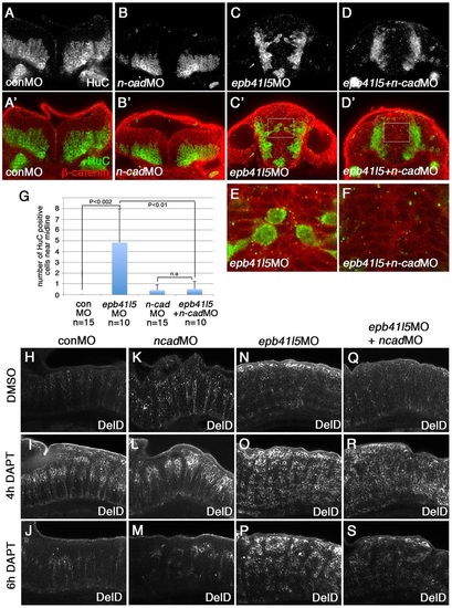
Reduced expression of n-cadherin rescues a delay of delamination and differentiation of neurons in epb41l5-deficient embryos. (A-F) Coronal sections showing localization of HuC-expressing neurons in the hindbrain. HuC-positive cells are only localized at the pial side of the hindbrain in control MO- or 0.1ng n-cadherin MO-injected embryos. A fraction of HuC-positive cells are mislocalized near the midline in epb41l5 morphants. The mislocalization of HuC-positive cells is rescued by partial knockdown of n-cadherin. The boxed areas in C′ and D′ are enlarged in E and F, respectively. (G) Statistical analysis of distribution of HuC-positive cells in the hindbrain. Error bars represent s.d. n.s., not significant. (H-S) DeltaD expression in the hindbrain. Lateral views. After 4h of DAPT treatment DeltaD expression is increased. After 8h of DAPT treatment, whereas DeltaD expression is decreased in control embryos and n-cadherin morphants, DeltaD expression persists in epb41l5 morphants. The persistent expression of DeltaD in epb41l5 morphants is rescued by partial knockdown of n-cadherin. |