
Fig. 3
- ID
- ZDB-FIG-160914-13
- Publication
- Bryan et al., 2016 - Loss of laminin alpha 1 results in multiple structural defects and divergent effects on adhesion during vertebrate optic cup morphogenesis
- Other Figures
- All Figure Page
- Back to All Figure Page
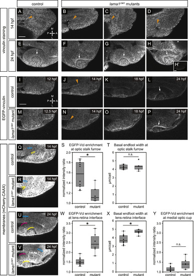
Focal adhesions are assembled in a specific spatiotemporal pattern, and are differentially disrupted by loss of lama1. (A-H) Antibody staining for the focal adhesion protein vinculin at 14 hpf (A-D) or 24 hpf (E-H), in control (A,E) or lama1UW1 mutant (B-D, F-H) embryos. (H’) No primary anti-vinculin antibody control. Orange arrowheads, location of optic stalk furrow; white arrows, lens-retina interface; dashed line, outline of optic vesicle. (I-P) Single confocal slices from 4D datasets of EGFP-vinculin (grayscale) localization during optic cup morphogenesis, ~12–24 hpf. (I-L) EGFP-vinculin recruitment in control embryo is apparent at the optic stalk furrow (J, orange arrowhead), and lens-retina interface (L, white arrow). (m-P) EGFP-vinculin recruitment in lama1UW1 mutant embryo at the optic stalk furrow (N, orange arrowhead), and lens-retina interface (P, white arrow). Dorsal views; scale bar, 50 µm. A, anterior; P, posterior; M, medial; L, lateral. (Q-Y) Quantification of EGFP-vinculin recruitment at the optic stalk furrow and lens-retina interface. (Q,R,U,V) mCherry-CAAX (membranes, gray) localization in same single confocal slices as J,N,L,P, respectively. At the optic stalk furrow or lens-retina interface (Q,R,U,V; yellow regions), EGFP-vinculin fluorescence intensity was normalized to mCherry-CAAX. Enrichment was measured by comparing to normalized EGFP-vinculin fluorescence intensity at the midline (Q,R,U,V; cyan regions). A control medial optic cup domain (U,V; magenta regions) was quantified as a location where EGFP-vinculin recruitment was not noted; this domain was also compared to normalized EGFP-vinculin fluorescence intensity at the midline (U,V; cyan regions). (S) Quantification of EGFP-vinculin recruitment indicates decreased ECM adhesion at the optic stalk furrow in the lama1UW1 mutant. (T) Average basal endfoot width at the optic stalk furrow, based on counting the number of cells within the quantified region; no significant difference indicates that differences in cell morphology at that position (e.g. basal constriction) cannot strictly account for differences in apparent EGFP-vinculin recruitment. (W) Quantification of EGFP-vinculin recruitment indicates increased ECM adhesion at the lens-retina interface in the lama1UW1 mutant. (X) Average basal endfoot width at the lens-retina interface, based on counting the number of cells within the quantified region; lama1UW1 mutants have fewer cells with wider basal surfaces, indicating that increased cell number or constriction cannot account for apparent increased EGFP-vinculin recruitment at the mutant basal surface. (Y) Quantification of EGFP-vinculin recruitment at a control site in the medial optic cup where no enrichment of vinculin recruitment occurred in control or lama1UW1 mutants. Quantifications were performed on 10 control embryos and 6 mutant embryos (one eye scored per embryo); n.s., not significant;*P<0.005, using a two-tailed t-test of unequal variance. |
| Antibody: | |
|---|---|
| Fish: | |
| Anatomical Terms: | |
| Stage Range: | 14-19 somites to Prim-5 |
| Fish: | |
|---|---|
| Observed In: | |
| Stage Range: | 14-19 somites to Prim-5 |
Reprinted from Developmental Biology, 416(2), Bryan, C.D., Chien, C.B., Kwan, K.M., Loss of laminin alpha 1 results in multiple structural defects and divergent effects on adhesion during vertebrate optic cup morphogenesis, 324-37, Copyright (2016) with permission from Elsevier. Full text @ Dev. Biol.