
Fig. 2
- ID
- ZDB-FIG-160914-12
- Publication
- Bryan et al., 2016 - Loss of laminin alpha 1 results in multiple structural defects and divergent effects on adhesion during vertebrate optic cup morphogenesis
- Other Figures
- All Figure Page
- Back to All Figure Page
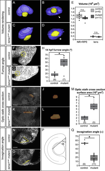
Quantitative analysis of lama1UW1 mutant phenotype. (A-E) Analysis of eye size at 24 hpf. (A-D) Volume renderings of control (A,B) and mutant (C,D) eyes. Neural retina + RPE (blue), lens (yellow). (A,C) Dorsal views. (B,D) Lateral views. arrowhead, choroid fissure; asterisk, choroid fissure missing in mutant. (E) Quantification of optic cup and lens volume in control and mutant eyes shows no significant difference in eye size. (F-H) Quantification of furrow angle during initial stages of optic stalk constriction. Single confocal section of control (F) and mutant (G) embryos, membrane channel (gray) at 14 hpf. A circle with a 25 µm radius was placed with its center at the vertex of the furrow, and angle was calculated by drawing radii to positions at which the circle intersected the optic vesicle and brain neuroepithelium. (H) Quantification of furrow angle demonstrates that the furrow exhibits a significantly larger angle in lama1UW1 mutants. (I-M) Visualization and quantification of optic stalk constriction. (I-L) 3D rendering of optic stalk cross-section (orange) over membrane channel (gray) at 24 hpf. (I,K) Dorsal views. (J,L) Face-on views of the optic stalk cross section. (M) Quantification of optic stalk cross section area shows that optic stalk constriction is impaired in lama1UW1 mutant embryos. (N-Q) Quantification of invagination angle. (N,O) Single confocal images of membrane channel (gray) in lama1UW1 control (N) and mutant (O) eyes at 24 hpf. Lines (yellow) were drawn to determine angle of invagination. (P) Schematic demonstrating how invagination angle (±) was determined. (Q) Quantification of invagination angle shows a severe defect in invagination in lama1UW1 mutant embryos. numbers at base of graph show embryos scored (one eye each);*P<0.001, using the student′s t-test. |
Reprinted from Developmental Biology, 416(2), Bryan, C.D., Chien, C.B., Kwan, K.M., Loss of laminin alpha 1 results in multiple structural defects and divergent effects on adhesion during vertebrate optic cup morphogenesis, 324-37, Copyright (2016) with permission from Elsevier. Full text @ Dev. Biol.