
Fig. 1
- ID
- ZDB-FIG-160914-11
- Publication
- Bryan et al., 2016 - Loss of laminin alpha 1 results in multiple structural defects and divergent effects on adhesion during vertebrate optic cup morphogenesis
- Other Figures
- All Figure Page
- Back to All Figure Page
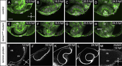
Timelapse confocal microscopy reveals severe defects in optic cup formation in lama1UW1 mutant. (A-H) Single confocal slices from 4D datasets of optic vesicle morphogenesis, 12.5-24.5 hpf. EGFP-CAAX (membranes, green), H2A.F/Z-mCherry (nuclei, magenta). (A-D) Optic cup formation in control embryo. (E-H) Optic cup formation in lama1UW1 mutant embryo. Dorsal views; scale bar, 50 µm. (I-M) Localization of laminin protein. (I-L) In control embryos, laminin protein is found lining basal surfaces of developing eye and brain. (M) lama1UW1 mutant embryo reveals absence of laminin protein from early stages of optic vesicle development. nk, neural keel; ov, optic vesicle; ec, ectoderm; br, brain; RPE, retinal pigmented epithelium; nr, neural retina; le, lens. A, anterior; P, posterior; M, medial; L, lateral. |
| Antibody: | |
|---|---|
| Fish: | |
| Anatomical Terms: | |
| Stage Range: | 5-9 somites to Prim-5 |
| Fish: | |
|---|---|
| Observed In: | |
| Stage Range: | 5-9 somites to Prim-5 |
Reprinted from Developmental Biology, 416(2), Bryan, C.D., Chien, C.B., Kwan, K.M., Loss of laminin alpha 1 results in multiple structural defects and divergent effects on adhesion during vertebrate optic cup morphogenesis, 324-37, Copyright (2016) with permission from Elsevier. Full text @ Dev. Biol.