
Fig. S1
- ID
- ZDB-FIG-160906-9
- Publication
- Pipalia et al., 2016 - Cellular dynamics of regeneration reveals role of two distinct Pax7 stem cell populations in larval zebrafish muscle repair
- Other Figures
- All Figure Page
- Back to All Figure Page
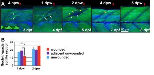
Cellular changes in wounded muscle. A. Lateral confocal image stack projections of wounded epaxial somites (red arrowhead) in larvae wounded at3 dpf and analysed 4 hours post wounding (hpw) or 1-5 dpw. Fixed larvae were stained for F-actin (Phalloidin) and nuclei (Hoechst). Note the broken fibres (asterisks) and rounded nuclei at the 4 hpw wound site. Loss of regular striation and presence of actin aggregates at 1 dpw (arrows) is followed by accumulation of nuclei in the wound site at 2 dpw (arrowheads). At 2 dpw, striations begin to reappear in thin fibres (yellow arrowheads) at the wound edge, and are abundant by 4 dpw. Dorsal to top, onterior to lcft. Bar = 50 µm. B. Quantification of nuclei per confocal transverse cross-section of epaxial somites that were wounded (red), adjacent to wound but unwounded (purple) and in unwounded fish (blue). Half the nuclei at the horizontal myoseptum were attributed to the epaxial somite. |