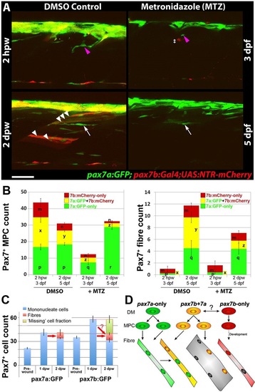
Ablation of pax7b-expressing cells diminishes marked fibres. pax7a:GFP;pax7b:NTR-mCherry larvae were treated overnight with MTZ or DMSO vehicle, then wounded and imaged by confocal 3D scanning at the indicated times. (A) Representative time-lapse images of wounded epaxial somites, showing MPC (magenta arrowheads) invasion of wound at 2hpw, followed by formation of nascent fibres at 2dpw (white arrows). Numerous large dual-labelled fibres are observed in control (white arrowheads). Anterior to left, dorsal to top. Scale bar: 50µm. (B) Counts of numbers (mean±s.e.m.) of red, green and dual-labelled MPCs and fibres in single wounded epaxial somites of control and MTZ-treated larvae. Counts omitted fast-moving phagocyte (‡ in A) and transient fibre labelling, which appeared rapidly only after wounding in MTZ-treated NTR-mCherry larvae and reflect the abundant release of mCherry from dying cells and uptake into injured fibres or phagocytes. Counts of each labelled cell type were compared between DMSO- and MTZ-treated larvae at 2hpw and 2dpw by two way ANOVA (with Bonferroni post hoc test, n=4 DMSO and 5 MTZ; for complete dataset and statistical analyses see Table S1). Within each cell type, distinct letters on columns (m,n,p,q,r,x,y,z) show groups that differed statistically at P<0.05. (C) Quantification (mean±s.e.m.) of mononucleate cells in epaxial wounded somites of pax7a:GFP or pax7b:GFP larvae reveals an increase at 1dpw, followed by a decrease at 2dpw, accompanied by formation of labelled fibres. Note the similar number of labelled fibres in each line (red arrows), and the apparent substantial reduction of labelled cells (‘missing’ fraction) only in the pax7b:GFP reporter line. (D) Modified founder cell model illustrating the known and hypothesised (?) behaviour of the pax7a:GFP-only MPC population (green), which contribute to wound repair and form nascent fibres, and the pax7b MPC population (yellow), which is more abundant, fuses to pre-existing (damaged?) fibres in the region of the wound and contributes to nascent fibre growth. We hypothesise that pax7a-only cells are founders initiating new fibre formation, whereas pax7b cells are FCMs contributing to fibre growth both in wounding and normal development. pax7b:RFP-only MPCs (red) form fibres early in development, accumulate at HZM and might act as a stem cell.
|
