
Fig. 6
- ID
- ZDB-FIG-160906-7
- Publication
- Pipalia et al., 2016 - Cellular dynamics of regeneration reveals role of two distinct Pax7 stem cell populations in larval zebrafish muscle repair
- Other Figures
- All Figure Page
- Back to All Figure Page
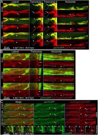
Fusion of pax7a- and pax7b-reporter cells to existing myotubes during wound repair. Extended orthogonal projection views of an epaxial somite wound in a pax7a:GFP;pax7b:gal4;UAS:RFP 4dpf larva showing individual pax7a:GFP;pax7b:RFP dual-labelled (yellow) MPCs fusing to existing unlabelled (A) or RFP+ (B) fibres from Movies 2 and 3. The whole image was non-linearly enhanced and brightness corrected to compensate for bleaching and facilitate tracking of individual cells, as described in Materials and Methods. (A) At 25.5hpw, prior to fusion, an MPC had uniform cytoplasmic GFP and diffuse cytoplasmic and vesicular RFP (arrow). 10min later, cytoplasmic GFP and RFP have now filled the whole cytoplasm of a large adjacent previously unlabelled fibre (brackets), whereas the vesicular RFP remains localised (arrowhead) and integrates into the fibre in the succeeding 50min (see Movie 2, blue MPC). Transverse II shows the same view as Transvere I, but with the fusing fibre marked (dots). (B) At 39.5hpw, two MPCs (magenta and yellow arrows), fuse simultaneously to the same large adjacent myotube (arrowheads; Movie 2, magenta and yellow MPCs). (C) At 32.3hpw, a dual-labelled MPC (arrowhead; Movie 3, white MPC) that originated from the anterior somite border, divided and then migrated along a recently formed GFP+ nascent myofibre (arrow). 10min later the MPC has fused into the nascent fibre, as shown by RFP loss from the MPC and increase in the fibre. The fused cell remains distinct at 10min, but merges into the nascent fibre by 20min. Process shown in merge and single colour lateral (dorsal up, anterior left) and transverse (dorsal up, medial left) views. Blue lines indicate range of extended orthogonal projection views. |
| Genes: | |
|---|---|
| Fish: | |
| Condition: | |
| Anatomical Terms: | |
| Stage Range: | Day 4 to Day 5 |