
Fig. 7
- ID
- ZDB-FIG-160826-17
- Publication
- Ji et al., 2016 - Mutations in zebrafish pitx2 model congenital malformations in Axenfeld-Rieger syndrome but do not disrupt left-right placement of visceral organs
- Other Figures
- All Figure Page
- Back to All Figure Page
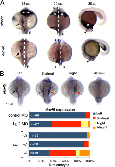
Asymmetric expression of elovl6 depends on Kupffer′s vesicle. (A) RNA in situ hybridizations revealed similar asymmetric expression patterns for pitx2c and evolv6 in left lateral plate mesoderm (black arrows) and brain (arrowheads) at 18-20 somite stages (ss). pitx2c mRNA was detected in hatching gland (asterisks) and neurons (white arrows), whereas elovl6 was not expressed in these domains. White dashed line is the midline; L=left and R=right. (B) Representative images are shown of left-sided, bilateral, right-sided and absent elovl6 expression in lateral plate mesoderm (arrows) at 18 somite stage. The graph shows quantification of asymmetric elovl6 expression profiles in control, Lgl2 depleted and genotyped cftrpd1048 mutant embryos. |
| Genes: | |
|---|---|
| Fish: | |
| Knockdown Reagent: | |
| Anatomical Terms: | |
| Stage Range: | 14-19 somites to 20-25 somites |
| Fish: | |
|---|---|
| Knockdown Reagent: | |
| Observed In: | |
| Stage: | 14-19 somites |
Reprinted from Developmental Biology, 416(1), Ji, Y., Buel, S.M., Amack, J.D., Mutations in zebrafish pitx2 model congenital malformations in Axenfeld-Rieger syndrome but do not disrupt left-right placement of visceral organs, 69-81, Copyright (2016) with permission from Elsevier. Full text @ Dev. Biol.