
Fig. 5
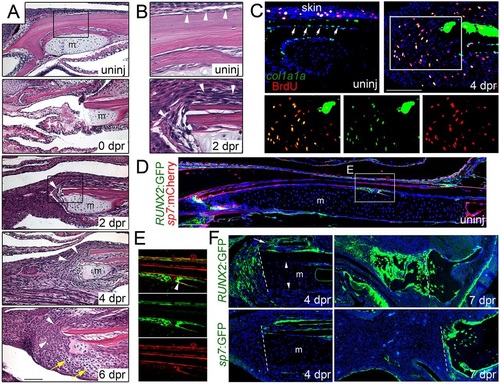
Mobilization of the periosteum in response to jaw resection. (A,B) H&E staining shows the posterior site of jaw resection before and during the first week of regeneration. In the uninjured animal, the lower jawbone is lined by a thin layer of periosteum (arrowheads, see inset B). Immediately after resection (0dpr), a small portion of Meckel′s cartilage (m) remains and soft tissue collapses into the section. By 2dpr, the periosteum (arrowheads) thickens and covers the cut surface of the bone (see inset B). The periosteum continues to expand into the resected region by 4dpr, and 2 days later, mesenchymal cells are seen throughout the resected area and early chondrocytes can be distinguished (yellow arrows). (C) Fluorescent in situ hybridization for col1a1a (green) combined with BrdU staining (red) shows col1a1a+ cells lining the uninjured jawbone periosteum (arrows), as well as the skin. At 4dpr, col1a1a expression increases in the periosteum and many col1a1a+ cells are seen in the mesenchyme within the resection zone. While col1a1a+ cells within the uninjured periosteum are largely negative for BrdU (in contrast to those within the skin), many BrdU+, col1a1a+ cells are seen in the mesenchyme near the resection site (see insets for magnified images, merged and single channels). (D) Section of an uninjured adult jaw shows sp7:mCherry (detected by anti-mCherry antibody) in osteoblasts lining bone and RUNX2:GFP (detected by anti-GFP antibody) in sparse patches of periosteum. (E) Magnification of boxed region in D shows RUNX2:GFP expression in periosteal cells underneath sp7:mCherry+ osteoblasts. A few cells co-express both transgenes (arrowhead), consistent with early differentiating osteoblasts. (F) After resection, RUNX2:GFP+ cells are found in the periosteum (arrow) overlying the jawbone and in expanding mesenchyme at 4dpr, but not in Meckel′s cartilage or its associated periochondrium (arrowheads). By 7dpr, both mesenchymal and early chondrocytes express RUNX2:GFP. By contrast, sp7:GFP labels the periosteum but not mesenchymal cells or chondrocytes at 4 and 7dpr. Hoechst labels nuclei in blue. Dashed lines indicate resection sites. Scale bars: 100µm. |