
Fig. 1
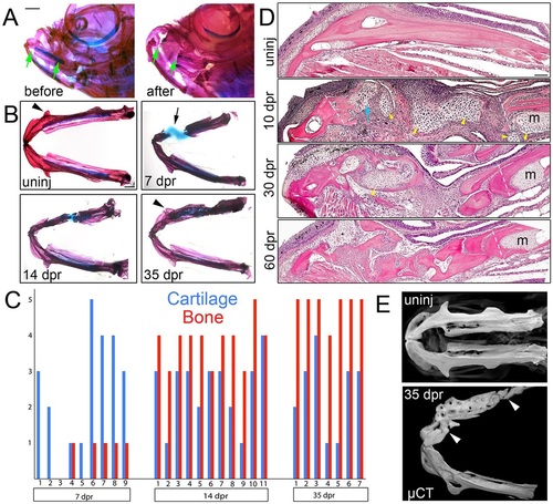
Regeneration of the lower jawbone in adult zebrafish. (A) Whole-mount views of adult zebrafish heads before and after resection stained with Alizarin Red and Alcian Blue to label bone and cartilage. Arrows show resection sites. (B) Dissected lower jaws show the time course of cartilage and bone formation during regeneration. Arrow denotes the repair cartilage that has contracted somewhat during mounting. Arrowheads indicate regeneration of the anguloarticular prominence. (C) Qualitative assessment of cartilage and bone formation during lower jaw regeneration in individual animals. The y-axis shows the amount of cartilage/bone in the lesion from none (0) to full spanning (5). (D) H&E staining on sections of the lower jawbone before and after resection. An extensive cartilage callus (yellow arrowheads) is seen at 10dpr, including at the anterior cut site (blue arrow) devoid of Meckel′s cartilage (m). Dashed lines show resection sites. (E) Bone µCT images show ventral views of lower jawbones from un-injured (uninj) and regenerated animals. Arrowheads indicate resection sites. See also Movie 1. Scale bars: 1mm in A,B and 100µm in D. |