
Fig. S6
- ID
- ZDB-FIG-160706-20
- Publication
- Raman et al., 2016 - aPKC regulates apical localization of Lgl to restrict elongation of microridges in developing zebrafish epidermis
- Other Figures
- All Figure Page
- Back to All Figure Page
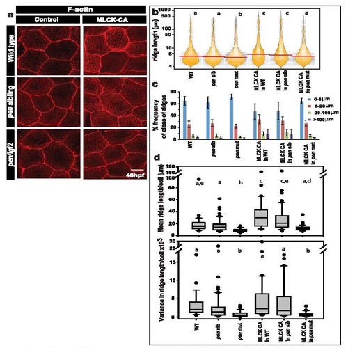
Over-expression of MLCK-CA does not give rise to longer ridges in pen/lgl2 mutants. Confocal microscopy analysis of the peridermal cells in given genotypes in either uninjected (control) or MLCK-CA injected embryos stained with phalloidin (a). Graphical representation of the distribution of ridge lengths and corresponding medians in various genotypes using bean plot (b). A bar graph showing percentage frequency distribution of ridges in short (0-5 µm), intermediate (5-20 µm), long (20-100 µm) and very long (>100 µm) categories (c). Representation of cell wise means and variances of ridge lengths in various genotypes using box-whisker plot (d). In (b) and (d) the distributions sharing the same alphabet do not differ significantly (Dunn’s multiple comparisons test, p-value < 0.05). Error bars in (c) are for the standard deviation. Scale bar in (a) is equivalent to 10 µm. |