
Fig. 6
- ID
- ZDB-FIG-160525-30
- Publication
- Zhao et al., 2016 - An essential role for Grk2 in Hedgehog signalling downstream of Smoothened
- Other Figures
- All Figure Page
- Back to All Figure Page
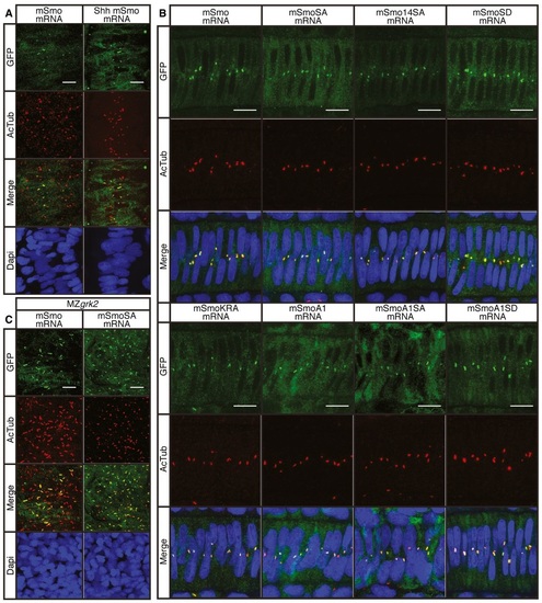
Cilia localisation of wildtype and mutant Smo A. Wildtype 18hpf embryos injected with mRNA encoding GFPtagged mSmo (green) showing localisation to the PC of myotomal cells labelled with antiacetylated tubulin (AcTub; red), stimulated in response to Shh injection (n = 4). Differences in PC distribution are due to morphological changes in the myotome induced by ectopic expression of Shh as revealed by the distribution of nuclei (DAPI stained; blue). Scale bar, 10 µm. B. Notochord cells of wildtype embryos expressing GFPtagged wildtype and mutant forms of mSmo. Note the localisation to the PC (labelled with antiAcTub; red) in each case (n = 4 for each sample). Scale bar, 10 µm. C. MZgrk2 18hpf embryos injected with mRNA encoding GFPtagged wildtype mSmo or mSmoSA (green) showing localisation to the PC (labelled with antiAcTub; red) in myotomal cells. More Smo is localised to the PC in MZgrk2 mutants compared to wild type (panel A). Scale bar, 10 µm. |