
FIGURE
Fig. 4
- ID
- ZDB-FIG-160512-11
- Publication
- Perner et al., 2016 - Analysis of Zebrafish Kidney Development with Time-lapse Imaging Using a Dissecting Microscope Equipped for Optical Sectioning
- Other Figures
- All Figure Page
- Back to All Figure Page
Fig. 4
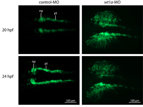
Knockdown of wt1a Disrupts Embryonic Kidney Development. Representative, extended depth of focus images from time-lapse recordings. In control morpholino injected embryos, kidney development shows normal progress with growing tubules and nephron primordia which start to fuse at the midline. In contrast, wt1a morphants fail to form proper nephron primordia and a massive amount of GFP positive cells are outside of the pronephric field. (np, nephron primordium; pt, pronephric tubule). |
Expression Data
| Gene: | |
|---|---|
| Fish: | |
| Knockdown Reagent: | |
| Anatomical Terms: | |
| Stage Range: | 20-25 somites to Prim-5 |
Expression Detail
Antibody Labeling
Phenotype Data
| Fish: | |
|---|---|
| Knockdown Reagent: | |
| Observed In: | |
| Stage Range: | 20-25 somites to Prim-5 |
Phenotype Detail
Acknowledgments
This image is the copyrighted work of the attributed author or publisher, and
ZFIN has permission only to display this image to its users.
Additional permissions should be obtained from the applicable author or publisher of the image.
Full text @ J. Vis. Exp.