
FIGURE
Fig. 4
- ID
- ZDB-FIG-160406-2
- Publication
- Roxo-Rosa et al., 2015 - The zebrafish Kupffer's vesicle as a model system for the molecular mechanisms by which the lack of Polycystin-2 leads to stimulation of CFTR
- Other Figures
- All Figure Page
- Back to All Figure Page
Fig. 4
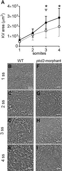
KV inflation live-dynamics. (A) KV luminal area of pkd2-morphants and WT embryos measured along development from 1 to 4 s.s. For each time point mean ±s.d. are indicated. Number of tested embryos: WT, n=11; pkd2-morphant, n=8. ψP<0.05, significantly different from the previous time point in pkd2-morphants; *P<0.05, significantly different from WT at the corresponding time point. (B-I) Bright field images captured for the same embryo along development are shown for the most representative WT (B-E) and pkd2-morphant (F-I). Scale bars: 10μm. |
Expression Data
Expression Detail
Antibody Labeling
Phenotype Data
| Fish: | |
|---|---|
| Knockdown Reagents: | |
| Observed In: | |
| Stage: | 1-4 somites |
Phenotype Detail
Acknowledgments
This image is the copyrighted work of the attributed author or publisher, and
ZFIN has permission only to display this image to its users.
Additional permissions should be obtained from the applicable author or publisher of the image.
Full text @ Biol. Open