
Fig. 5
- ID
- ZDB-FIG-160329-5
- Publication
- Bojarczuk et al., 2016 - Cryptococcus neoformans Intracellular Proliferation and Capsule Size Determines Early Macrophage Control of Infection
- Other Figures
- All Figure Page
- Back to All Figure Page
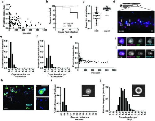
Polysaccharide capsule is smaller on intracellular cryptococci and is greatly enlarged after infection.(a) Association between inoculum and proportion of intracellular cryptococci. Each point is a separate infection. P-value 0.0499. 120 infections of Tg(fms:Gal4.VP16)i186 ; Tg(UAS:nfsB.mCherry)i149 zebrafish with C. neoformans strain H99GFP. (b) Survival of AB strain zebrafish infected with >102-103 C. neoformans strain H99 or mutant cap59. P < 0.0001, logrank (Mantel-Cox). hazard ratio = 10.1 (logrank; 95% confidence interval 5.6, 18.4). 89 and 81 infections from H99 and cap59 groups respectively, from n = 8. (c) Proportion of intracellular cryptococci 4 hours post infection of Tg(mpeg1:mCherryCAAX)sh378 with >101-102C. neoformans strain H99 or mutant cap59 labelled with Calcofluor white. Quantitation of 22 infected fish from n = 3 experiments. (d-f) In vivo measurement of intracellular and extracellular polysaccharide capsule radius. 1025 cryptococci were measured from 50 infections from n = 5 repeats. (d) Maximum intensity projection from three dimensional fluorescence imaging of Tg(mpeg1:mCherryCAAX)sh378 (magenta), that labels macrophage membranes, infected with inocula between >101-102 of C. neoformans strain H99 labeled for polysaccharide capsule (green) and cell wall (cyan). Boxed areas are enlarged in i and ii, and are single z-sections. Scale bar is 20 µm in left image and 5 µm in i and ii. (e,f) Intracellular cryptococci have smaller polysaccharide capsules. Relative frequency histograms of capsule radius for intracellular (e) and extracellular (f) cryptococci. (g) Output of probability model using relative numbers within macrophages from (e,f) to calculate proportion of intracellular cryptococci at different inocula given random input (h) Cryptococcal capsule is enlarged and shed at 24 hpi. Fixed tissue of AB strain zebrafish infected with >102-103 C. neoformans strain H99GFP (green) at 24 hpi labeled with antibody to capsular polysaccharide (cyan). Scale bar 10 µm. Boxed areas are enlarged in i and ii. Scale bar 5 µm. (i,j) Cryptococcal capsule size greatly enlarged 24 hpi. Relative frequency histograms of capsule radius for inoculum (i) and 24 hpi (j) cryptococci isolated from AB strain zebrafish infected with >102-103 of C. neoformans strain H99. Inset panels are example India ink stained samples. Scale bar 5 µm. 615 cryptococci were measured in 12 infections from n = 3. |