
Fig. S4
- ID
- ZDB-FIG-160309-19
- Publication
- Tang et al., 2016 - Imaging tumour cell heterogeneity following cell transplantation into optically clear immune-deficient zebrafish
- Other Figures
- All Figure Page
- Back to All Figure Page
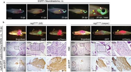
Visualising neuroblastoma and ERMS metastasis in transplanted rag2E450fs (casper) fish. (a) Retro-orbital transplantation of a GFP-labelled neuroblastoma. White arrow indicates the site of injection; yellow arrow indicates metastasis to a region near the liver. (b) Intra-muscular transplantation of ERMS into rag2E450fs recipient fish. White arrow indicates the site of injection; yellow arrows indicate the site of metastasis. Note that metastasis can be directly visualised by epifluorescence microscopy in the rag2E450fs (casper) recipients (yellow arrow) but not the rag2E450fs (AB) (open yellow arrow). H&E and anti-GFP staining on sections of the recipient animals confirmed the location of metastasis. Scale bars equal 5 mm for whole animal images, 2 mm in images of heads, 1 mm in 40x histological images; 300 µm in 100x histological images; and 100 µm in 400x histological images. |