
FIGURE
Fig. S1
- ID
- ZDB-FIG-160309-16
- Publication
- Tang et al., 2016 - Imaging tumour cell heterogeneity following cell transplantation into optically clear immune-deficient zebrafish
- Other Figures
- All Figure Page
- Back to All Figure Page
Fig. S1
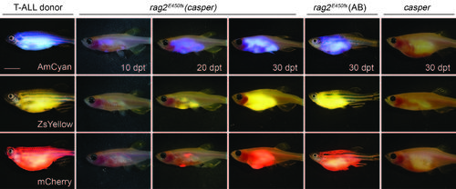
Imaging T-ALL progression and growth in transplanted rag2E450fs (casper) fish. Monoclonal T-ALLs were serially passaged in CG1 strain fish and then used as donors (left panel). Cells were transplanted intra-peritoneally into rag2E450fs (casper), rag2E450fs (AB), and unconditioned, casper-strain recipient fish (1.0x10⁵ cells per recipient animal). Merged brightfield and fluorescent images are shown at 10, 20 and 30 dpt. Scale bars equal 5 mm. |
Expression Data
Expression Detail
Antibody Labeling
Phenotype Data
Phenotype Detail
Acknowledgments
This image is the copyrighted work of the attributed author or publisher, and
ZFIN has permission only to display this image to its users.
Additional permissions should be obtained from the applicable author or publisher of the image.
Full text @ Nat. Commun.