
Fig. 4
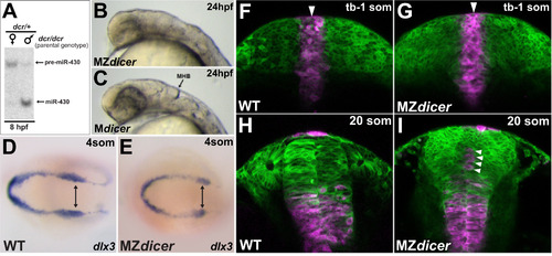
miR-430 activity is required post-gastrulation within proximal neural progenitors (A) Northern blot analysis of miR-430 expression: lane 1, embryos derived from dcr/dcr female germline (maternally deficient) X dcr/+ male; lane 2, embryos derived from dcr/dcr male germline X dcr/+ female (maternally sufficient). Embryos deficient for maternal dicer function do not generate significant levels of mature miR-430 until after 8hpf. (B) MZdicer embryo (maternally and zygotically deficient) fails to generate contiguous neural lumen and ventricular organization. (C) Mdicer embryo (maternally deficient) undergoes ventricle formation, suggesting that zygotic Dicer contribution (and delayed miR-430 production) is sufficient for neural tube generation. (D and E) In situ analysis of dlx3 expression, marking the lateral edges of neural keel. (F–I) Kaede lineage tracing after photoconversion at dorsal midline (tailbud-1somite stage; F, G) and ~10 hours later (20 somite stage; H, I). Arrowheads demarcate dorsal midline. As ingression proceeds, proximal progenitors adopt ventral positions in neural rod. In MZdicer embryos, a subset of proximal progenitors fails to ingress ventrally (arrowheads in panel I). |
Reprinted from Developmental Biology, 409(2), Takacs, C.M., Giraldez, A.J., miR-430 regulates oriented cell division during neural tube development in zebrafish, 442-50, Copyright (2016) with permission from Elsevier. Full text @ Dev. Biol.