
Fig. 1
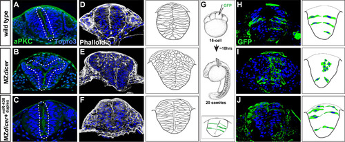
miR-430 is required for neural tube morphogenesis. Transverse sections through the hindbrain region of 20-somite stage embryos. Topro3, nuclear stain. Wildtype neural progenitors (A, D and H) display apical localization of aPKC and bilateral organization along the presumptive neural midline (A), display normal subcellular localization of F-actin (D), and acquire elongated cell morphology (H). In contrast, MZdicer neural progenitors (B, E and I) form ectopic apical membranes (B), display abnormal F-action organization (E), and fail to establish contact with lateral membrane and acquire epithelial character in dorsal regions (I). (C, F and J) Neural progenitors from MZdicer embryos injected at the 1-cell stage with miR-430 RNA duplex display rescued cell morphology and organization. (G) Scatter labeling strategy. 1-2 blastomeres of 16-cell embryos were injected with GFP mRNA. |
Reprinted from Developmental Biology, 409(2), Takacs, C.M., Giraldez, A.J., miR-430 regulates oriented cell division during neural tube development in zebrafish, 442-50, Copyright (2016) with permission from Elsevier. Full text @ Dev. Biol.