
Fig. 5
- ID
- ZDB-FIG-160219-41
- Publication
- Duncan et al., 2016 - Hypothalamic radial glia function as self-renewing neural progenitors in the absence of Wnt/ß-catenin signaling
- Other Figures
- All Figure Page
- Back to All Figure Page
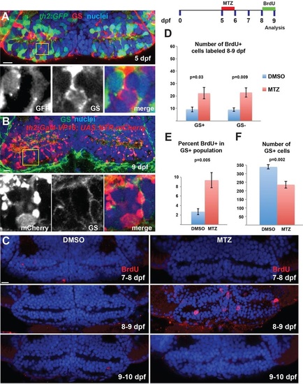
Hypothalamic radial glia proliferate in response to ablation of dopaminergic progeny. (A,B) The th2:GFP (5dpf, A) and th2:Gal4 (9dpf, B) transgenes do not label GS+ radial glia. Yellow boxes indicate regions shown beneath. (C) Ablation of th2:Gal4+ cells from 5-6dpf leads to increased BrdU labeling only at 8-9dpf. (D,E) Ablation of th2:Gal4+ cells increases the number and percentage of GS+ radial glia, as well as the number of GS cells, labeled with BrdU from 8-9dpf. (F) The overall number of GS+ radial glia is decreased at 9dpf following ablation of th2:Gal4+ cells. Images are single optical sections from ventral views of whole-mount brains. Error bars indicate s.e.m.; n=50 optical sections from five brains for each experiment. Scale bars: 10µm. |