
Fig. 1
- ID
- ZDB-FIG-160219-37
- Publication
- Duncan et al., 2016 - Hypothalamic radial glia function as self-renewing neural progenitors in the absence of Wnt/ß-catenin signaling
- Other Figures
- All Figure Page
- Back to All Figure Page
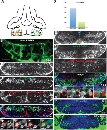
Radial glia in the hypothalamic posterior recess are not Wnt-responsive. (A) Diagram of the hypothalamic posterior recess from a ventral view of a 5dpf zebrafish brain. Radial glia are in green; the red box indicates the area depicted in B. (B) High-magnification optical section of her4.3:EGFP expression, showing the position of cell soma and radial processes. (C) Radial glial cells in the 5dpf posterior recess can be labeled with anti-Glutamine synthetase (GS, blue), a her4.3:EGFP transgene (green), and a Gal4 enhancer trap line (red). Yellow box indicates the region shown beneath. A triple-labeled cell is indicated by red circles. (D) Most cells expressing GS or her4.3:EGFP also express the other marker. Error bars indicate s.e.m.; n=22 optical sections from two brains. (E) Most GS+ and her4.3:EGFP+ radial glia do not express the Wnt reporter transgene 7xTCF-Xla.Siam:GFP. Yellow box indicates region shown beneath. A reporter-negative radial glial cell is indicated by green circles, and a reporter-expressing non-glial cell is indicated by red circles. (F) GS+ cells are not reduced in number in lef1 mutants at 5dpf. Images are single optical sections from ventral views of whole-mount brains. WT, wild type; nuclei are stained with Hoechst 33342 (blue). Scale bars: 10µm. |