
FIGURE
Fig. 5
- ID
- ZDB-FIG-151221-6
- Publication
- Chua et al., 2015 - Tumor-specific signaling to p53 is mimicked by Mdm2 inactivation in zebrafish: insights from mdm2 and mdm4 mutant zebrafish
- Other Figures
- All Figure Page
- Back to All Figure Page
Fig. 5
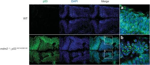
mdm2-/-; p53M214K/M214K embryos accumulate mutant p53 protein. Whole mount immunohistochemistry was performed on mdm2-/-; p53M214K/M214K and wild-type embryos at 30 hpf. The embryos were fixed in methanol:acetone (1:1) and stained with p53-5.1 hybridoma supernatant (green) and DAPI counterstain (blue). The stained embryos were deyolked, dorsally mounted in glycerol and imaged at the midbrain at × 40 magnification using a confocal microscope. (a, b) Magnified insets showing different mutant p53 localization in different cells in the mdm2-/-; p53M214K/M214K embryo. |
Expression Data
| Gene: | |
|---|---|
| Antibody: | |
| Fish: | |
| Anatomical Term: | |
| Stage: | Prim-15 |
Expression Detail
Antibody Labeling
Phenotype Data
| Fish: | |
|---|---|
| Observed In: | |
| Stage: | Prim-15 |
Phenotype Detail
Acknowledgments
This image is the copyrighted work of the attributed author or publisher, and
ZFIN has permission only to display this image to its users.
Additional permissions should be obtained from the applicable author or publisher of the image.
Full text @ Oncogene