
Fig. S5
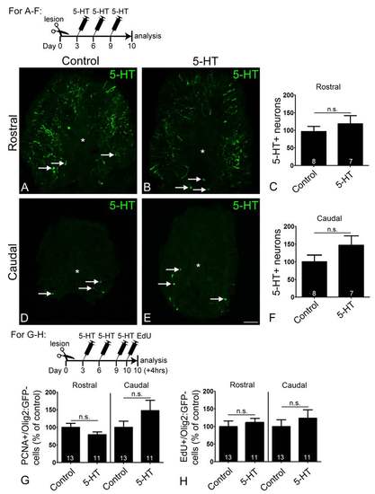
Serotonin (5-HT) injections do not increase numbers of serotonergic neurons or proliferation of ventricular cells outside the pMNlike domain. A-F: Cross sections through the spinal cord are shown; asterisks indicate the central canal; arrows indicate serotonergic neurons. Serotonin injections do not significantly increase numbers of serotonergic neurons rostral (Mann-Whitney U-test, P = 0.5358) or caudal (Mann-Whitney U-test, P = 0.2810) to the lesion site. G,H: In contrast to the cells in the olig2:GFP+ pMN-like domain (compare Fig. 4F-I), the number of PCNA+ (Student’s t-test, Rostral P = 0.1640, Caudal P = 0.1656) or EdU+ (Student’s t-test, Rostral P = 0.5525, Caudal P = 0.4323) ventricular cells outside that domain (open arrows in Fig. 4F,G), were not significantly altered by addition of serotonin. Scale bar in E = 50 µm. |