
Fig. S4
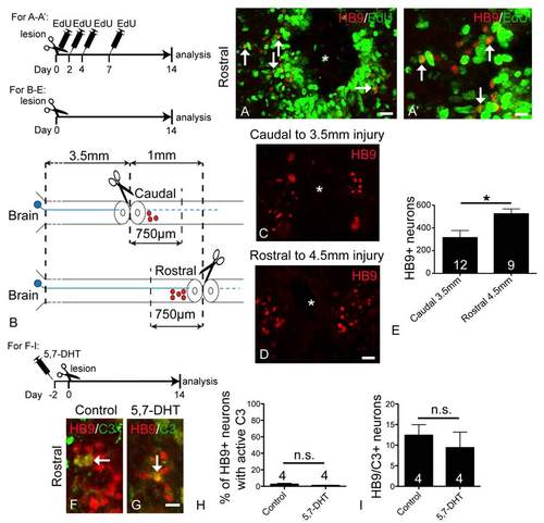
Evidence that HB9 immuno-reactive motor neurons are newly generated after a lesion (A, A′), that descending axons influence generation of motor neurons (B-E) and that serotonin signaling does not affect cell death of regenerated motor neurons (F-I). A,A′: Cross sections through the spinal cord are shown; asterisks indicate the central canal. A′ is a higher magnification of the left middle area in A. Multiple EdU injections (see timeline) lead to double labeling of almost half of the newly present HB9+ neurons (arrows) after a lesion. B-E: Placing lesions 1 mm apart makes it possible to compare neurogenesis in the same intervening stretch of spinal cord with either descending axons present (caudal lesion) or absent (rostral lesion) (B). Labeling (C,D) and counts (E) of new HB9+ motor neurons in these equivalent areas up to 750 µm away from the lesion site indicated significantly more new HB9+ motor neurons when the lesion was situated caudal to sampled area (Student’s t-test, *P = 0.0184). F-I: Double labeling with HB9 and caspase 3 antibodies in control animals and those in which descending serotonergic axons were ablated, did not show increased proportions (Mann-Whitney U-test, P = 0.3429) or numbers (Mann-Whitney Utest, P = 0.6857) of double-labeled cells (arrows) rostral to the lesion site at 14 days post-lesion. Bar in A = 25 µm; in A′ = 12.5 µm; in D = 25 µm for C,D; in G = 12.5 µm for F,G. |