
Fig. 5
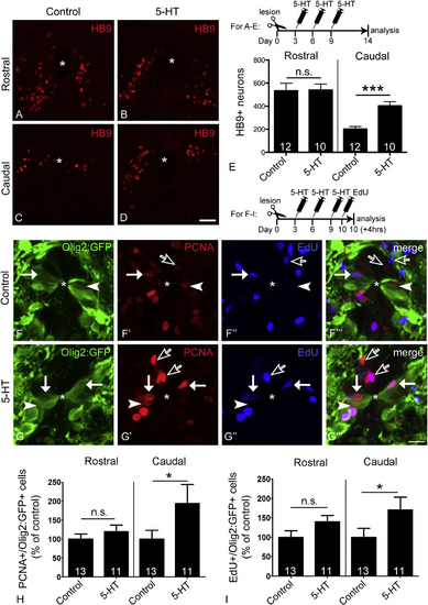
Serotonin Injections Increase the Number of Newly Generated Motor Neurons and Proliferating pMN-like Progenitor Cells Caudal to the Spinal Lesion Cross-sections through the spinal cord are shown; asterisks indicate the central canal, arrowheads indicate PCNA+/olig2:GFP+ cells, solid arrows indicate EdU+/olig2:GFP+ cells, and empty arrows indicate PCNA+/olig2:GFP or EdU+/olig2:GFP cells. (A–E) Serotonin injection doubles the number of newly generated motor neurons caudal to the lesion but has no effect rostral to the lesion (see timeline in E for experimental paradigm; Student’s t test; p < 0.0001). (F–I) In the ventricular zone of olig2:GFP transgenic animals, the numbers of PCNA+/olig2:GFP+ (Mann-Whitney U-test; p = 0.0437) and EdU+/olig2:GFP+ (Mann-Whitney U-test; p = 0.0231) pMN-like ERGs are significantly increased only caudal to the lesion (see timeline for experimental paradigm). The scale bar in (D) represents 50 µm for (A)–(D) and in (G′′′) represents 10 µm for (F) and (G). See also Figure S5. |