
Fig. 2
- ID
- ZDB-FIG-151123-1
- Publication
- Liang et al., 2015 - Kaempferol Identified by Zebrafish Assay and Fine Fractionations Strategy from Dysosma versipellis Inhibits Angiogenesis through VEGF and FGF Pathways
- Other Figures
- All Figure Page
- Back to All Figure Page
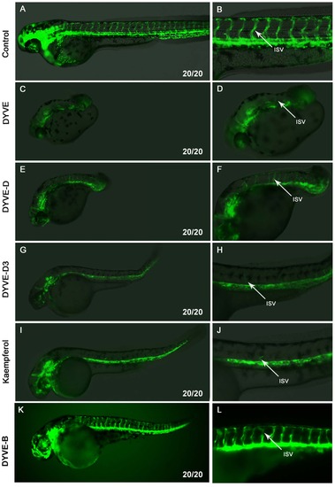
Fraction DYVE-D3 and kaempferol inhibited the outgrowth of ISVs. Embryos at 48 hpf anterior is to left and dorsal is to top. Right column is enlargement of left column. (A,B), control group, ISV fully developed. (C,D) DYVE group, embryonic trunk formation was impaired and the head was small, while the ISVs were still present. (E,F) DYVE-D group, the embryonic trunk was very short, and ISVs developed. (G,H) DYVE-D3 group, the embryo developed slower than the control, but did not have strong defects. Outgrowth of ISV was inhibited. (I,J) kaempferol group, embryos developed slower than control, but were mostly normal. Outgrowth of ISVs was inhibited. (K,L) DYVE-B group, the embryos were slightly abnormal on the morphology, but ISVs appeared normal. White arrows point to ISV. Phenotype ratio is showed in the lower right corner. The working concentration of DYVE, DYVE-D, DYVE-D3 and DYVE-B was based on Supplementary Table S1, and kaempferol group was 40 µM. |