
Fig. 4
- ID
- ZDB-FIG-151119-8
- Publication
- Bhumika et al., 2015 - Decreased thyroid hormone signaling accelerates the reinnervation of the optic tectum following optic nerve crush in adult zebrafish
- Other Figures
- All Figure Page
- Back to All Figure Page
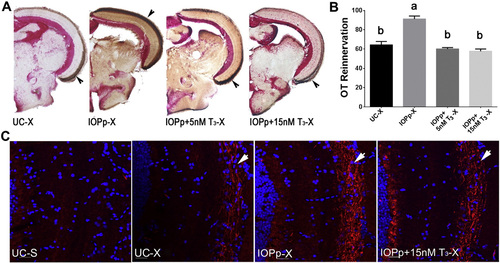
T3 supplementation attenuates OT reinnervation. A. Reinnervation of the OT by RGC axons at 7 dpi in different groups. Brown staining indicated with arrow represents biocytin labeled RGC axons. B. Quantification of the area covered by RGC axons in the OT of the different groups. Data represent mean ± SEM (n = 4–5 animals/group). Data were analyzed by ANOVA followed by Scheffé test. Means with a different letter (a–b) are significantly different (p at least < 0.05). C. Immunofluorescent detection of Gap43 protein (red) in RGC axons (arrow) arriving in the OT. Cell nuclei are stained with DAPI (blue). Pictures are representative images for n = 4–5 animals/group. Scale bar — 20 µm. For all panels: UC—untreated control, IOPp—iopanoic acid pretreated for 2 weeks, IOPp + 5nM T3 (or + 15 nM T3)-iopanoic acid pretreated for 2 weeks before crush and co-treated with 5 nM T3 (or 15 nM T3) starting 3 days before crush, S—sham operated, X—optic nerve crushed, OT—optic tectum. |
Reprinted from Molecular and cellular neurosciences, 68, Bhumika, S., Lemmens, K., Vancamp, P., Moons, L., Darras, V.M., Decreased thyroid hormone signaling accelerates the reinnervation of the optic tectum following optic nerve crush in adult zebrafish, 92-102, Copyright (2015) with permission from Elsevier. Full text @ Mol. Cell Neurosci.